Micromate™ Case Report series presents real cases performed by the physicians currently using our full-fledged robotic platform for percutaneous procedures.
Today, we join Dr. Alexander Kupferthaler, Interventional Radiologist at the Ordensklinikum Linz – Barmherzige Schwestern, for a robot-guided bone biopsy.
Clinical Context
65-year-old female with invasive lobular carcinoma of the breast with skeletal carcinomatosis.
After years of progression-free survival, a PET-CT scan detected several novel, abnormal metabolic bone lesions. A bone biopsy of the iliac crest was selected for accessibility.
Interventional Procedure
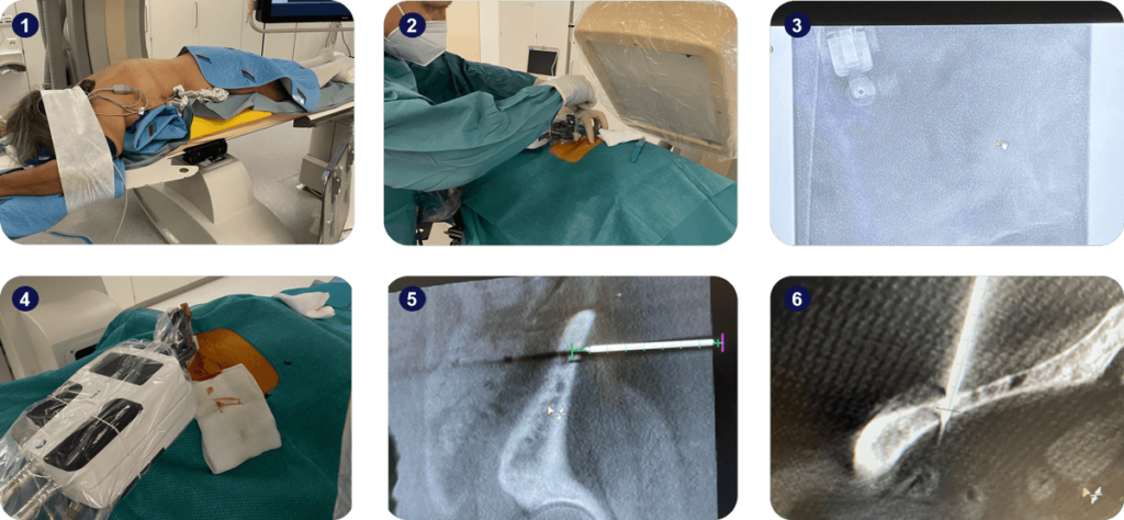
Image fusion and planning were performed using a Philips Allura Xper FD20 angiography device and Xper Guide planning software. Two needles with different trajectories were planned for a biopsy of the same lesion. The target point location was defined halfway through the left iliac crest. The patient was positioned prone using iFIX for comfortable arm and head positioning, and the left side of the pelvis was lifted prior to the sterile field creation.
Micromate™ was gross-positioned near the entry point of the first surgical plan and aligned to the trajectory within 60 seconds under fluoroscopic live imaging. After alignment, a 13G needle was manually inserted until a stable bone purchase was achieved and a control scan was acquired. The biopsy needle was drilled through the bone for tissue harvesting using the Teleflex Arrow OnControl system, also under live imaging. The fine alignment procedure was repeated for the second trajectory without the need for adjusting the gross position.
Micromate™ reached the desired target accurately in both cases and ensured stable guidance while taking the biopsy. A 0.07mm accuracy on the Entry Point view and a 0.77-degree angular accuracy along the trajectory in Progress View were achieved for the first trajectory. For the second tissue harvesting, Entry Point view accuracy was not reported, and a 1.02 degrees angular accuracy was achieved.

Key Takeaway
Micromate™ enabled the clinical team to insert multiple needles within the range of motion of the robot, without the need to manually reposition the holding arm.
Adequate patient positioning using iFIX allowed an accurate and safe robotic execution of the surgical trajectory.


