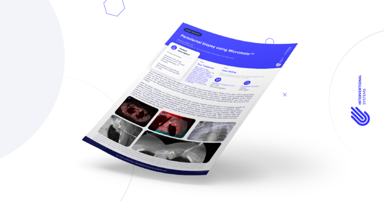
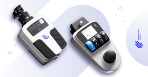
Micromate™ Case Report series presents real cases performed by the physicians currently using our full-fledged robotic platform for percutaneous procedures.
For this case report, we join Dr. Marco van Strijen, Interventional Radiologist at St. Antonius Ziekenhuis – Nieuwegein, the Netherlands, for a robot-assisted parasternal biopsy.
Clinical Context
The patient was a 54-year-old female diagnosed with breast carcinoma in 2017. She was treated with chemotherapy and reached remission.
A follow-up PET-CT scan detected paracardial and parasternal lesions.
Interventional Procedure
An intra-operative 3D scan of the patient in supine position was performed using a Philips Allura Xper FD20 angiography device, and fused with diagnostic PET-CT.
The suspicious lesion was segmented, and the surgical trajectory planned using the Xper Guide planning software. Then, a target point for the insertion of the biopsy needle was defined at the distal border of the segmented lesion, to ensure tissue harvesting covered the whole lesion.
Micromate™ was then gross positioned near the predefined entry point and remotely controlled for alignment to the surgical plan under fluoroscopic live imaging.
After the robotic alignment, an 17G tru-cut biopsy needle was coaxially inserted twice through a 17G guiding needle for tissue harvesting.
The whole procedure lasted 19 minutes and a (breast) metastatic adenocarcinoma was diagnosed. Post-operative accuracy measurements indicated a guidance needle accuracy of 0.00mm (i.e., placement of the needle within the 2 cm cut window) and an angular displacement of 1.41 degrees along the trajectory in the Progress View.
The post-operative 3D CBCT scan showed no pneumothorax. However, a follow-up conventional chest X-ray was taken 4 hours after the procedure, and pneumothorax signs have been found, requiring the insertion of a small chest drain. The coaxial guidance needle was accurately placed.
The complication is likely due to a more distal, deliberated, physician-determined insertion of the tru-cut biopsy needle than planned.

Key Takeaways
Micromate™ helped the clinical team standardize the workflow for percutaneous soft-tissue biopsies.
The stable instrument guidance of the system enabled the team to coaxially advance biopsy devices through a guidance needle, preventing harmful angular deviations during insertion caused by surrounding tissue or breathing.
Case Rating*
Radiation exposure: 48% less radiation (6.57 mSv)
Procedure duration: 33% faster (19 min)


