Micromate™ Case Report series presents real cases performed by the physicians currently using our full-fledged robotic platform for percutaneous procedures.
For this case report, we head out to St. Antonius Hospital, in the Netherlands, to join Dr. Marco van Strijen for the cryoablation of an osteoid osteoma of the tibia.
Clinical Context
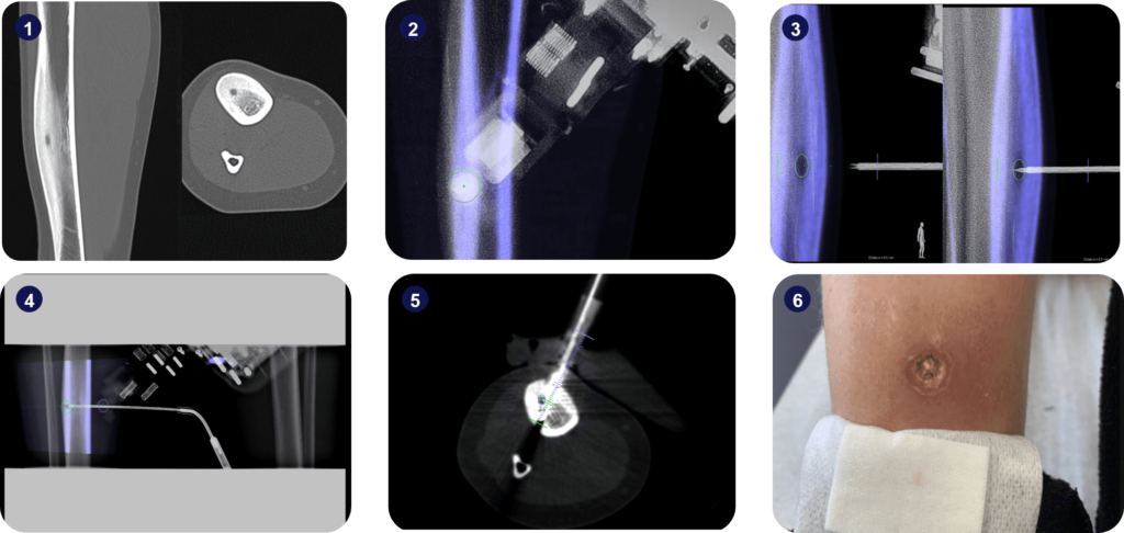
The patient was a 9-year-old female who’d exhibited swelling of the right ventral tibia for a few years and reported pain in the months prior to the intervention.
Diagnostic X-ray and CT scans suggested the existence of an osteoid osteoma.
Interventional Procedure
An intra-operative 3D scan of the patient’s leg was performed in supine position using a Philips Allura Xper FD20 angiography device.
The suspicious lesion was segmented, and the surgical trajectory was planned using the Xper Guide planning software. A target point was defined at the distal border of the segmented lesion and an intermediate target point was marked in the center of the lesion.
Micromate™ was gross-positioned near the entry point and aligned to the trajectory under live fluoroscopic imaging.
After alignment, an 11G needle was advanced until the intermediate target point, using a Teleflex Arrow OnControl Bone Drill System, and a control scan was acquired. This allowed the creation of a path for the insertion of the cryoablation probe.
The probe (Boston Scientific IcePearl) was advanced towards the intermediate target point. A control scan was acquired, and the probe further advanced towards the target point. Cryoablation was then performed with IcePearl at 40%.

Postoperative measurements of the guidance needle’s final location indicated a trajectory alignment accuracy of 0.00mm on the Entry Point View, a tip location accuracy of 0.00mm in the Progress View, and an angular displacement of 0.52 degrees along the trajectory in the Progress View.
The physician-controlled cryoablation needle tip location accuracy was 1.30mm and the angular displacement along the trajectory was 1.21 degrees in the transversal view, and no tip location or angular inaccuracies have been detected in Progress View.
Key Takeaway
Micromate™ allowed the clinical team to perform a successful cryoablation in a case where instrument access and alignment stability would be challenging if performed freehand.
Case Rating*
Radiation exposure: 87% less radiation (1.68 mSv)


