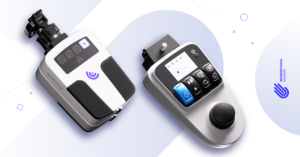
Micromate™ Case Report series presents real cases performed by the physicians currently using our easy-to-use miniature robot for percutaneous procedures.
For this case report, we join Dr. Marco van Strijen, Interventional Radiologist at St. Antonius Ziekenhuis – Nieuwegein, the Netherlands, for a robot-assisted percutaneous lung biopsy.
Clinical Context
The patient was a 52-year-old male referred for suspicion of rectal carcinoma.
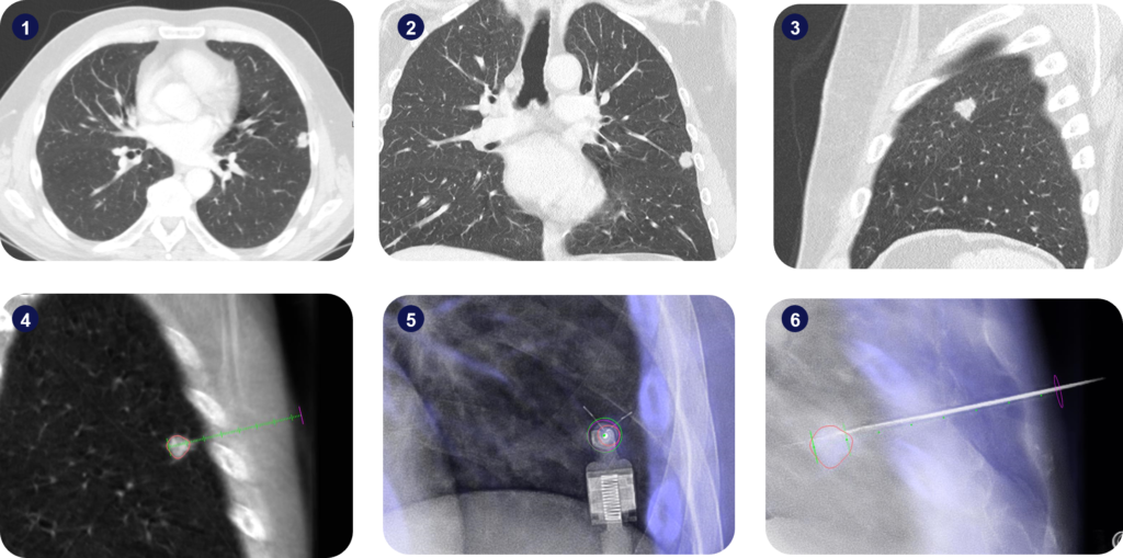
A CT scan was acquired for staging and a full lateral lesion has been identified in the left lung.
Interventional Procedure
An intra-operative 3D scan of the patient in supine position was performed using a Philips Allura Xper FD20 angiography device.
The suspicious lesion was segmented, and the surgical trajectory planned using the Xper Guide planning software. Then, an intermediate target point for inserting the guidance needle was defined at the proximal border of the segmented lesion, and a target point for the insertion of the biopsy needle was defined at the distal border of the segmented lesion, to ensure tissue harvesting covered the whole lesion.
Micromate™ was then gross-positioned near the predefined entry-point and remotely controlled for alignment to the surgical plan under fluoroscopic live imaging.
After the robotic alignment, an 18G biopsy needle was coaxially inserted three times through a 17G guiding needle for tissue harvesting.
A metastatic colon carcinoma was diagnosed. The procedure lasted 16 minutes and the patient had no complications. Post-operative accuracy measurements indicated a trajectory alignment accuracy of 0.56mm on the Entry Point View and an angular displacement of 1.48 degrees along the trajectory in the Progress View.

Key Takeaway
Micromate™ allowed the clinical team to accurately target a lung lesion in the vicinity of the pleura under live imaging, without complications such as pneumothorax.
Case Rating*
Radiation exposure: 70% less radiation (3.79 mSv)
Procedure duration: 43% faster (16 min)


