Micromate™ Case Report series presents real cases performed by the physicians currently using the world’s smallest robot for percutaneous procedures.
Today, we join Drs. Gerlig Widmann and Martin Freund, Interventional Radiologists at the Tirol Kliniken, for a robot-guided type II endoleak embolization.
Clinical Context
Patient’s profile: Male patient, previously subjected to an aorta endovascular aneurysm repair (AEVAR), diagnosed with an 18cm deep type II endoleak, with severe enlargement of the aneurysmal sac.
Interventional Procedure
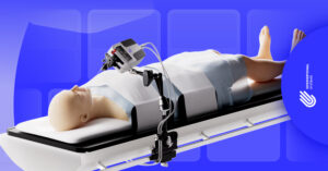
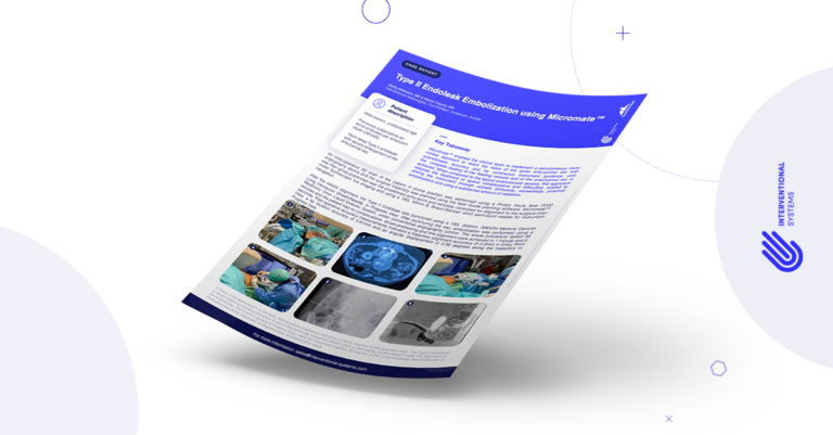
An intra-operative 3D scan of the patient in prone position was performed using a Philips Allura Xper FD20 angiography device and the surgical trajectory was planned using the Xper Guide planning software. Micromate™ was then gross-positioned near the predefined entry-point and remotely controlled for alignment to the surgical plan under fluoroscopic live imaging and using an 18G, 40mm B. Braun-Sterican short semi-blunt needle for instrument navigation.
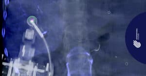
After the robotic alignment, the type II endoleak was punctured using a 19G, 200mm, ARGON Medical Devices Long Introducer Sheath/Needle, in Progress View. After puncturing the sac, embolization was performed using a Medtronic Onyx Liquid Embolic System, under conventional angiography control. The whole procedure lasted 49 minutes and the patient had no complications. Gross-positioning and fine alignment were achieved in 1 minute and 5 seconds. Post-operative accuracy measurements indicated a trajectory alignment accuracy of 0.0mm in Entry Point View, a tip location accuracy of 0.81mm, and an angular displacement of 0.39 degrees along the trajectory in Progress View.
Key Takeaway
Micromate™ enabled the clinical team to implement a percutaneous trans-lumbar approach to reach the nidus of the aortic aneurysmal sac with submillimeter accuracy and for continuous instrument guidance until the complete sealing of the feeding vessels and of the aneurysmal sac is achieved. When compared to the typical endovascular access, this approach reduces the likelihood of typical complications and difficulties related to driving the instrument through vessels (tortuosity, exceedingly proximal embolization) and to using a substantial amount of radiation.