Micromate™ Case Report series presents real cases performed by the physicians currently using the world’s smallest robot for percutaneous procedures.
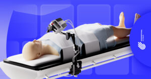
For this report, we’re following Dr. Marco van Strijen, Interventional Radiologist at the St. Antonius Hospital, during a robot-guided para-aortic biopsy.
Clinical Context
The patient was a 54-year old female with Non-Hodgkin lymphoma under treatment; PET+ para-aortic lymph node as a new finding. Differential diagnosis included metastatic breast disease, differentiating lymphoma or granuloma.
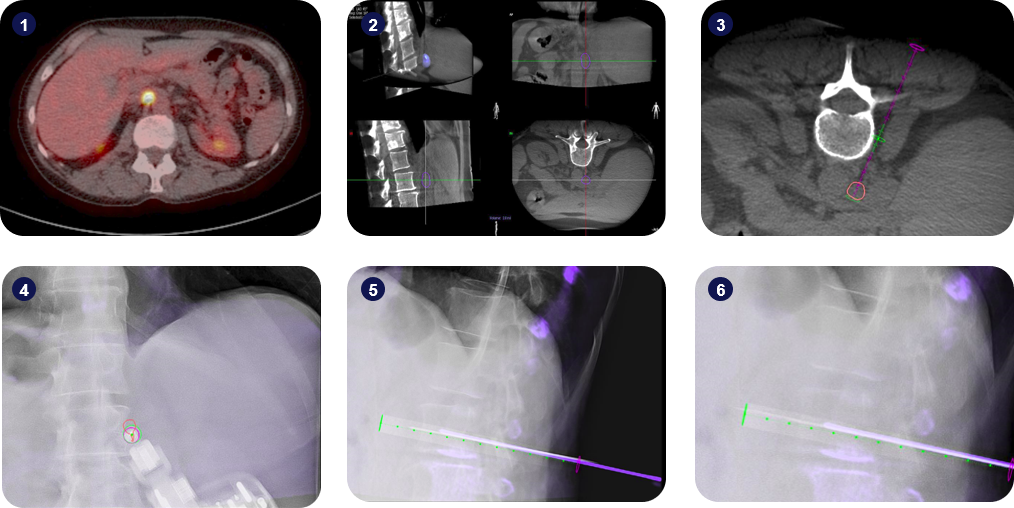
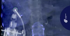
1) Fusion of the pre-operative PET+ and CT scans, clearly highlighting the new lesion on the para-aortic lymph node; 2) Lesion segmentation and visualization of the patient’s anatomy during planning; 3) Definition of the surgical plan. The red contour corresponds to the borders of the segmented lesion; 4) Alignment of Micromate™ to the surgical plan under live imaging; 5-6) Insertion of the guidance needle and tissue harvesting.
Interventional Procedure
An intra-operative 3D scan of the patient in supine position was performed using a Philips Allura Xper FD20 angiography device. The suspicious lesion was segmented, and the surgical trajectory planned using the Xper Guide planning software. An intermediate target point has been defined along the trajectory for the placement of a guidance needle, and a target point for the insertion of the biopsy needle has been defined at the distal border of the segmented lesion, to ensure tissue harvesting covered the whole lesion.
Micromate™ was then gross-positioned near the predefined entry-point and remotely controlled for alignment to the surgical plan under fluoroscopic live imaging. After the robotic alignment, an 18G biopsy needle was coaxially inserted twice through a 17G guiding needle for tissue harvesting. A large B-cell lymphoma was diagnosed, in concordance with previous findings in the bone marrow.
The procedure lasted 26 minutes and the patient had no complications. Post-operative accuracy measurements indicated a trajectory alignment accuracy of 0.0mm on the Entry Point View and an angular displacement of 1.0 degrees along the trajectory in the Progress View.
Key Takeaways
Micromate™ helped the clinical team achieve submillimeter accuracy during a biopsy on a delicate anatomy, essential for an accurate diagnosis.
Micromate™ is self-locking after alignment to the surgical plan, retaining an accurate positioning for instrument guidance during the whole procedure. This enabled the clinical team to coaxially insert multiple instruments with clinically optimal accuracy.